Micro Fest
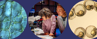
Join us for a celebration of seeing small through a variety of tools, from simple hand lenses to powerful microscopes. Uncover textures, patterns, and ecosystems that are normally invisible to the naked eye. Meet drifting plankton, explore immune cells, identify parasites, and dive into hands-on activities that invite you to think small—and see even smaller.
1:00–3:00 p.m.: Meet the artists in Gallery 1.
Featuring award-winning images and video reels from Nikon’s renowned photomicrography competition, this digital showcase is sure to amaze you.
Enjoy a curated selection of photos and videos from past contests, revealing cells, crystals, insects, and plant structures in astonishing detail. Some of them were captured by artists from the Bay Area, who will share their inspiration and process in Osher Gallery 1 from 1 to 3 p.m.
Nikon Small World is regarded as the leading forum for showcasing the beauty and complexity of life as seen through the light microscope. The Photomicrography Competition is open to anyone with an interest in microscopy and photography. The video competition, entitled Small World In Motion, encompasses any movie or digital time-lapse photography taken through the microscope.
With Dr. Steven Ruzin
The largest public collection of historic microscopes in the world, the Golub Collection contains antique microscopes that span from the seventeenth through the twentieth century. Take a look at rare microscopes that represent huge leaps in microscopy. Learn from the collection’s curator Dr. Steven Ruzin, and get an exciting glimpse into the incredible history of microscopy.
The Golub Collection was established in 1994 when Dr. Orville J Golub gave his initial donation of 32 antique microscopes to the University of California, Berkeley. Since then, the collection has grown through further Golub donations and curator purchases to include nearly 200 microscopes. It is on permanent display in the Onderdonk Lobby of the Valley Life Sciences building on the UC Berkeley campus.
With the San Francisco Microscopical Society
Why do green algae glow bright red under a fluorescence microscope? How are tiny diatoms arranged into delicate displays? Answer these questions and more with the SF Microscopical Society and microscopists from Merritt College, who bring a variety of different microscopes and samples to explore.
Founded in 1870, the San Francisco Microscopical Society is a community of aspirational, amateur, and professional microscopists in the Bay Area. They welcome all people of any age or background, from those who have not used a microscope to those who have been working with them for decades.
With Nate Dube
Enjoy gorgeous, microscopic mosaics made from diatoms, and learn about algae’s enormous impact on the environment.
Diatoms are tiny but powerful—scientists estimate they produce between twenty and fifty percent of the oxygen we breathe. If that’s not enough, they’re also beautiful, showcasing a variety of delicate geometric shapes. Join artist and scientist Nate Dube to take a close look at diatom arrangements and marvel at their intricacies.
Nate Dube is the vice president of the San Francisco Microscopical Society. Their microscopy journey began with an interest in Victorian diatom arranging. They are constantly experimenting with new tools and techniques for creating and photographing slides, and hope to inspire curiosity and make the microscopic world more accessible to all.
With the Fletcher Lab
Explore life at the microscopic scale with the NTDscope, a mobile-based microscope developed in the Fletcher Lab. See familiar creatures up close and learn how these microscopes are used to diagnose diseases in remote areas
The Fletcher Lab at UC Berkeley is an interdisciplinary group of bioengineers, biophysicists, and molecular biologists developing tools and techniques to understand how cells move, communicate, and get sick.
With the Biohub Bioengineering Team
Join scientists and engineers from Biohub to explore the Remoscope, a portable device they developed to diagnose malaria. Remoscope uses embedded machine learning to inspect blood cells as they flow past a simple ultraviolet light microscope. It can find malaria parasites in infected blood faster and more consistently than a trained human—come see for yourself!
The Bioengineering Team at Biohub is a team of multidisciplinary engineers who build novel instrumentation ranging from laboratory automation and custom microscopes, to portable diagnostic equipment for global health.
With the Weiner Lab
Birds flock, fish school, and ants swarm—many organisms work in groups to solve critical tasks. Did you know your immune cells also work together in similar ways? Join us to watch these amazing immune cells move under the microscope as they swarm to sites of injury and infection. Get inside the mind of the cell to understand why this teamwork is so essential to keep you safe.
The Weiner Lab at UCSF seeks to understand how single cells and groups of cells decide where to go—from immune cells hunting bacteria, to skin cells healing wounds, to the complex cell movements that build a human being.
With the Weiner Lab
Cells are constantly sending messages—and you can too. Join scientists from the Weiner Lab and become part of a living network. Channel your inner cell, follow the rules of communication and movement, and watch pathways grow and shift across the floor, revealing how information spreads through a system and coordinates group motion.
The Weiner Lab at UCSF seeks to understand how single cells and groups of cells decide where to go—from immune cells hunting bacteria, to skin cells healing wounds, to the complex cell movements that build a human being.
With Dana Carrison-Stone and Hitachi
Take a very detailed look at your favorite (or least favorite) household insects! Exploratorium biologist Dana Carrison-Stone will demonstrate the use of a scanning electron microscope (SEM), which can take images at over 1000x magnification by shooting an electron ray at its subject. Admire the hairy legs of bees, see eye to eye with a spider, and find out if ants are actually cute—or just plain scary.
Dana Carrison-Stone is the Living Systems Lab manager here at the Exploratorium. She has a background in marine biology and a love for all things small and strange.
With Veronica Johnson
Phytoplankton may be small, but these microscopic algae have large impacts on the food web around them, from shellfish to birds to humans. Take a peek into their world and understand the important role they play in the ecosystem.
Veronica Johnson was a biologist in the Exploratorium's Bio Lab for nine years, where she fostered a phytoplankton fascination. She has spent many hours studying plankton sample collected from the Bay and is ready to share her enthusiasm with you.
With Point Blue Conservation Science
Zooplankton—small animals that drift through the sea—form the base of marine food webs, feeding everything from fish to whales. Their features and behavior also provide researchers with vital information on ocean health and environmental shifts.
Join scientists from Point Blue Science to dive deep into the lives of zooplankton. Peer through microscopes, play interactive games,, and discover the surprising diversity inside a single drop of seawater.
With 160 scientists, Point Blue Conservation Science works to reduce the impacts of climate change, habitat loss, and other environmental threats while developing nature-based solutions to benefit both wildlife and people.
With Peter Taylor
How can microscopes better serve museum guests? Come explore a work-in-progress microscope, the MicroCOSM, designed to meet the specific needs of museum visitors. Chat with the lead designer and get to know his thoughtful creation.
Parameter Explorations is a fledgling company building museum-duty microscopes. Their first product, the MicroCOSM, is almost ready. Get a peek into their design process!
Peter Taylor has designed science exhibits for museums for over a decade. He was frustrated by the limitations of the available microscope options he had to build exhibits around, so he decided to solve the problem. His design attempts to be modular, adjustable, configurable, all while maintaining excellent optics and ease of use and accessibility for the visitor.
With the Healthy Housing and Vector Control Mosquito Lab
Get to know one of the world’s most notorious insects in this hands-on exploration of mosquito biology. Observe live mosquito larvae under a stereoscope, use a pipette to take a closer look, and follow their transformation through each stage of metamorphosis. Learn how mosquitoes spread disease and how their life cycle shapes the world around us.
The Healthy Housing and Vector Control Mosquito Lab is a team of certified Vector Control Technicians with the San Francisco Department of Public Health. The lab conducts surveillance for health nuisances caused by rats and mosquitoes and surveillance for invasive mosquitoes as well as for West Nile Virus. In addition to fieldwork, the lab actively participates in and provides engaging educational programs for all ages to help the community understand causes and ways to prevent vector‑borne diseases.
With the Visitor Engagement Facilitators
Build your own magnification tools and create custom slides with everyday objects. What do they look like under close examination?
Grab a miniscope kit and convert your phone into a portable, picture-taking miniscope. Use it to see the Exploratorium at its smallest scale, and then take it home to continue your exploration of the ultra tiny!
While supplies last.
Limited capacity and recommended for families with kids ages 4 to 8.
Join us for Storytime Science and enjoy an engaging storybook read-aloud followed by a related activity geared toward young children (and their grown-ups). Come hear a story, get hands-on, and learn something new with the Exploratorium’s own Vivian Altmann!
Visit the Exploratorium at Pier 15


Supporters

This material is based upon work supported by the Center for Cellular Construction, which is a Science and Technology Center funded by the National Science Foundation. NSF STC Cooperative Agreement AWARD: DBI-1548297. Any opinions, findings, and conclusions or recommendations expressed in this material are those of the author(s) and do not necessarily reflect the views of the National Science Foundation.